|
|
Dr.
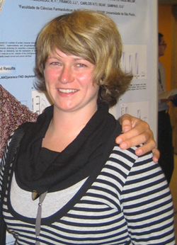
Lidija Kovačič
Department of Molecular and Biomedical Sciences email: lidija.kovacic PhD thesis summary
Ammodytoxins (Atxs) are presynaptically neurotoxic
(β-neurotoxic) secreted phospholipases A2
(sPLA2) from the long-nosed viper (Vipera a. ammodytes) venom. The exact molecular mechanism of
β-neurotoxicity is still unknown,
but it is clear that the toxicity of Atx is due to binding to the specific
receptors in nerve endings of the motoneurons and its enzymatic
activity. In neuronal tissues, the interaction of Atx with
intracellular binding proteins, calmodulin ( Using the
commercial cross-linking reagent sulfo-SBED and AtxC we synthesized a new
molecular tool for detection and characterization of Atx-binding proteins.
Using photo-reactive sulfo-SBED-AtxC, we detected some new Atx-binding
proteins in different tissues and cell lines, which escaped the detection
with previously used methods. We improved isolation procedures to obtain
sufficient amounts of active Atx-receptors (R25 and others) to further
characterize and identify them. In isolated fractions we found some known
neuronal proteins, however, their function as Atx-receptors should be
additionally confirmed. One of them was synaptotagmin I, which could well
represent a missing element in our understanding of the process of
β-neurotoxicity. Using sulfo-SBED-AtxC, we found that the toxin was
rapidly internalized into the differentiated PC12 cells in culture as it
interacted with CaM in the cytosol of these cells already within a few minutes.
On the other hand, using non-neuronal cells, sulfo-SBED-AtxC did not complex
and label CaM, probably because it could not reach CaM in such cells.
Translocation of Atx into the cytosol of cells is therefore rapid and likely
a neuro-specific process. By the means of sulfo-SBED-AtxC we mapped the
interaction surface between AtxC and its binding proteins, CaM, 14-3-3p and
PDI. Using computer modeling, we built three-dimensional (3D) models of the
respective complexes, which will be used to plan experiments to unravel the
role of these interactions in the process of β-neurotoxicity. In this
way, the 3D model of a complex between Atx and CaM explains very well
experimentally determined stabilization of Atx in cytosol-like conditions as
well as the up to 20-fold increase in the enzymatic activity of Atx when
complexed to CaM. We also constructed 3D models of some physiologically
important sPLA2s and the model-based predictions matched
experimental observations in showing that GV and GX sPLA2 in the
complex with CaM become substantially more enzymatically active. This
indicates an important role of CaM in regulation of enzymatic activity of
sPLA2s in the cytosol of cells. The mechanism of the observed
activation of Atx by CaM was kinetically dissected and found to be best
described as a nonessential activation, and probably represents one of the
crucial factors in expression of the ß-neurotoxicity in the case of Atxs. |
